In love with a new image from yesterday’s work on the focused ion beam/scanning electron microscope! The sample center looks somewhat like a morel mushroom, with blobs of silicate in a network of iron-rich melt. A particularly cheerful blob can be found in the upper right corner of the region of interest.
Composition map by energy-dispersive X-ray analysis in scanning electron microscopy (colors tweaked).
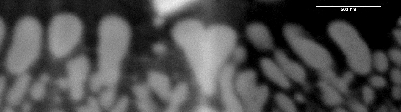
Scanning electron photomicrograph of region of silicate heart (the new header).
